
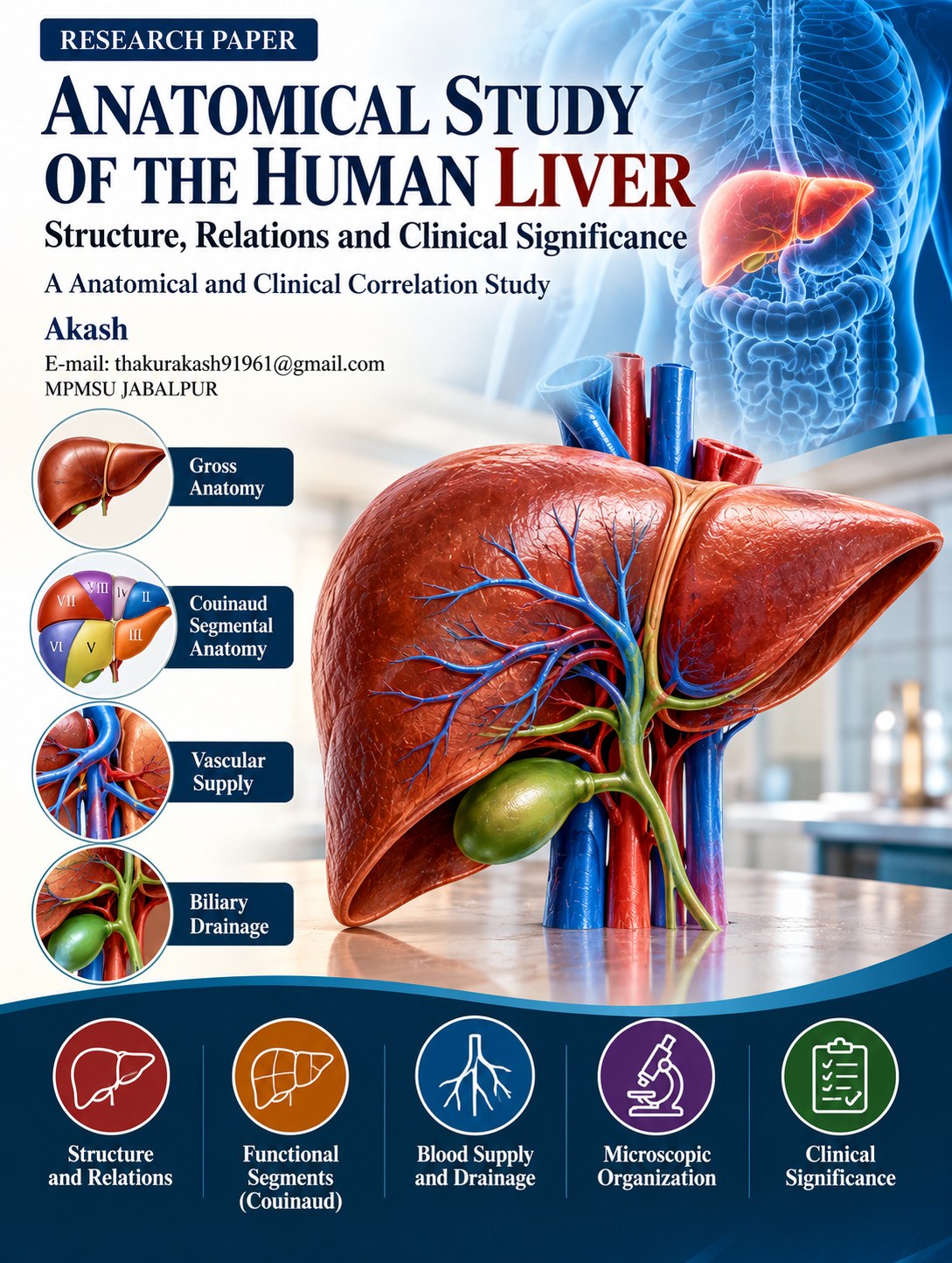
ANATOMICAL STUDY OF THE HUMAN LIVER Structure, Relations and Clinical Significance
Keywords:
Liver, anatomy, hepatic segments, Couinaud classification, portal vein, hepatic artery, liver testsAbstract
The liver is the largest internal gland and one of the most important organs of the human body. It occupies a major portion of the right upper quadrant of the abdominal cavity and has complex anatomical relationships with the diaphragm, stomach, duodenum, gallbladder, kidneys, inferior vena cava and major vascular structures. Its dual blood supply from the portal vein and hepatic artery, together with venous drainage through the hepatic veins, gives the liver a distinctive vascular organization.
Modern understanding of hepatic anatomy has progressed from the traditional morphological division into right and left lobes to functional segmental anatomy, particularly the Couinaud classification. This system divides the liver into eight functionally independent segments based on portal venous distribution and hepatic venous planes, providing a clinically useful framework for imaging, biopsy, resection and transplantation.
This review describes the gross anatomy, surfaces, borders, lobes, ligaments, vascular supply, biliary drainage, lymphatic drainage, nerve supply, microscopic organization and clinically important anatomical relationships of the human liver. It also correlates hepatic anatomy with laboratory investigations commonly used in liver assessment, including ALT, AST, alkaline phosphatase, bilirubin, albumin and PT/INR.
Downloads
References
1. Bloomston, M., & Reddy, S. K. (2010). Liver anatomy. Surgical Clinics of North America, 90(4), 643–653.
Source: https://pubmed.ncbi.nlm.nih.gov/20637938/
2. van den Esschert, J. W., de Graaf, W., van Lienden, K. P., Heger, M., & van Gulik, T. M. (2013). Hepatic anatomy. Seminars in Liver Disease, 33(3), 232–244.
Source: https://pmc.ncbi.nlm.nih.gov/articles/PMC6448666/
3. Bismuth, H. (1982). Surgical anatomy and anatomical surgery of the liver. World Journal of Surgery, 6, 3–9.
Source: https://pubmed.ncbi.nlm.nih.gov/7052609/
4. Mathew, R. P., & Venkatesh, S. K. (2018). Liver vascular anatomy: A refresher. Abdominal Radiology, 43(8), 1886–1895. https://doi.org/10.1007/s00261-018-1623-z
Source: https://pubmed.ncbi.nlm.nih.gov/29696320/
5. Covey, A. M., Brody, L. A., Maluccio, M. A., Getrajdman, G. I., & Brown, K. T. (2002). Variant hepatic arterial anatomy revisited: Digital subtraction angiography performed in 600 patients. Radiology, 224(2), 542–547.
Source: https://pubmed.ncbi.nlm.nih.gov/12147874/
6. Kwo, P. Y., Cohen, S. M., & Lim, J. K. (2017). ACG Clinical Guideline: Evaluation of abnormal liver chemistries. American Journal of Gastroenterology, 112(1), 18–35.
Source: https://pubmed.ncbi.nlm.nih.gov/27995906/
7. Kmieć, Z. (2001). Cooperation of liver cells in health and disease. Advances in Anatomy, Embryology and Cell Biology, 161, III–XIII, 1–151. https://doi.org/10.1007/978-3-642-56553-3
Source: https://pubmed.ncbi.nlm.nih.gov/11729749/
8. DeLeve, L. D. (2013). Liver sinusoidal endothelial cells and liver regeneration. Journal of Clinical Investigation, 123(5), 1861–1866.
Source: https://pubmed.ncbi.nlm.nih.gov/23635785/
9. Friedman, S. L. (2008). Hepatic stellate cells: Protean, multifunctional, and enigmatic cells of the liver. Physiological Reviews, 88(1), 125–172.
Source: https://pubmed.ncbi.nlm.nih.gov/18195085/
10. Wisse, E., Braet, F., Duimel, H., Geerts, W., Luo, D., & van der Smissen, P. (1996). Structure and function of sinusoidal lining cells in the liver. Hepatology, 24(1), 198–201.
Source: https://pubmed.ncbi.nlm.nih.gov/8707268/
11. Sutherland, F., & Harris, J. (2002). Claude Couinaud: A passion for the liver. Archives of Surgery, 137(11), 1305–1310.
Source: https://pubmed.ncbi.nlm.nih.gov/12437404/
12. Strasberg, S. M. (2005). Nomenclature of hepatic anatomy and resections: A review of the Brisbane 2000 system. Journal of Hepato-Biliary-Pancreatic Surgery, 12, 351–355.
Source: https://pubmed.ncbi.nlm.nih.gov/16133703/
13. Nagino, M., Kamiya, J., Uesaka, K., et al. (2006). Anatomic right hepatic trisectionectomy. Journal of the American College of Surgeons, 202(3), 462–468.
Source: https://pubmed.ncbi.nlm.nih.gov/16500251/
14. Kuntz, E., & Kuntz, H.-D. (2008). Hepatology: Principles and Practice. Springer.
Source: https://link.springer.com/book/10.1007/978-3-540-76839-7
15. Gebhardt, R., & Hovhannisyan, A. (2010). Organ-specific metabolic zonation in the liver. Pharmacology & Therapeutics, 127(1), 30–39.
Source: https://pubmed.ncbi.nlm.nih.gov/20430054/
16. Lautt, W. W. (2009). Regulatory processes interacting to maintain hepatic blood flow stability: The hepatic arterial buffer response. Hepatology, 49(6), 2080–2083.
Source: https://pubmed.ncbi.nlm.nih.gov/19475693/
17. Renz, J. F., & Busuttil, R. W. (2000). Current surgical therapy of liver tumors. Surgical Clinics of North America, 80(1), 103–119.
Source: https://pubmed.ncbi.nlm.nih.gov/10685149/
18. Kikuchi, S., et al. (2017). Clinical anatomy of the liver: Review of the 19th Meeting of the Japanese Research Society of Clinical Anatomy. Anatomical Science International, 92, 257–269.
Source: https://pmc.ncbi.nlm.nih.gov/articles/PMC5340221/
19. World Health Organization. (2024). Hepatitis. World Health Organization.
Source: https://www.who.int/health-topics/hepatitis
20. Kawada, N., et al. (2010). Hepatic stellate cells in liver fibrosis and regeneration. Journal of Gastroenterology and Hepatology, 25(Suppl. 1), S3–S9.
Downloads
Published
Issue
Section
License
Copyright (c) 2026 Akash bhilala (Author)

This work is licensed under a Creative Commons Attribution 4.0 International License.