Pharmacological Evaluation of the Wound Healing Potential of Trichosanthes dioica Leaf Extract Ointment in an Excision Wound Model in Wistar Rats
Keywords:
Trichosanthes dioica, wound healing, excision wound model, ointment, interleukin-6, histopathologyAbstract
Background: Wound healing is a complex, multi-phase biological process, and there is continuing interest in plant-derived agents that can accelerate repair while limiting excessive inflammation. Trichosanthes dioica Roxb., a plant traditionally credited with anti-inflammatory, antioxidant and tissue-regenerative properties, was evaluated for its wound healing potential in the present study.
Aim: To pharmacologically evaluate the wound healing properties of Trichosanthes dioica leaves in an experimental excision wound model.
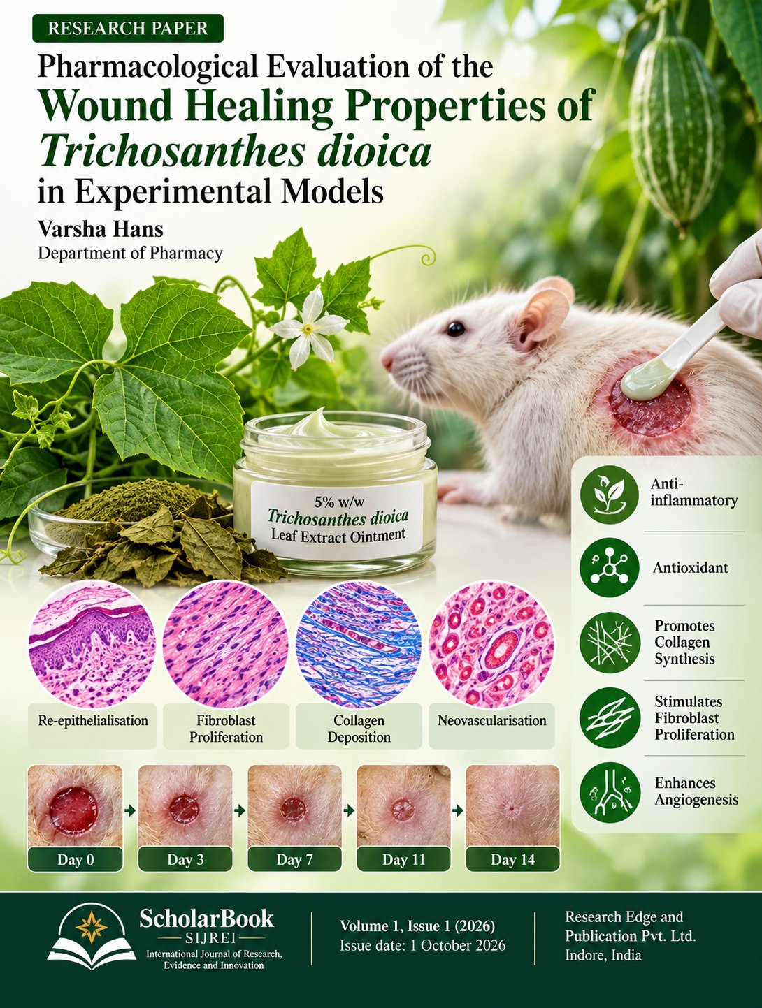
Methods: A 5% w/w ointment of the hydroalcoholic leaf extract of T. dioica, obtained by Soxhlet extraction, was formulated in a soft paraffin base. Eighteen male Wistar rats were divided into three groups (n = 6): an untreated control, a Plermin-treated standard group, and a group treated once daily with the 5% T. dioica ointment, following creation of a full-thickness circular excision wound. Wound contraction was recorded on Days 0, 3, 7, 11 and 14; on Day 14 animals were euthanised and granulation tissue was assessed histopathologically (H&E and Masson's Trichrome staining) and analysed for interleukin-6 (IL-6) by ELISA.
Results: The test ointment produced a progressive reduction in wound area that closely paralleled the standard group and was markedly faster than the untreated control, with wound contraction of 98.91% versus 56.83% (control) by Day 14. Histopathology of the test group showed enhanced re-epithelialisation, fibroblast proliferation, collagen deposition and neovascularisation compared with the control. Tissue IL-6 concentration was lowest in the test group, indicating attenuation of the inflammatory phase.
Conclusion: Topical application of 5% w/w Trichosanthes dioica leaf extract ointment significantly enhanced wound healing in rats, supporting its traditional use and warranting further phytochemical and clinical investigation.
Downloads
References
1. Khandaker, M., Akter, S., & Imam, M. Z. (2018). Trichosanthes dioica Roxb.: A vegetable with diverse pharmacological properties. Food Science and Human Wellness, 7(1), 34–48. https://doi.org/10.1016/j.fshw.2017.12.005
2. Zilani, M. N. H., Uddin, S. J., Hossain, H., Hazni, H., Shilpi, J. A., & Hossain, M. G. (2018). Chemical characterization and bioactivity of Trichosanthes dioica edible shoot extract. Oriental Pharmacy and Experimental Medicine, 18(2), 167–175. https://doi.org/10.1007/s13596-018-0310-5
3. Bhattacharya, S., Haldar, P. K., & Ghosh, A. K. (2010). Paralytic and lethal effects of Trichosanthes dioica root extracts in experimental worms. Pharmaceutical Biology, 48(9), 960–965. https://doi.org/10.3109/13880200903420847
4. Harborne, J. B. (1998). Phytochemical methods: A guide to modern techniques of plant analysis (3rd ed.). Chapman & Hall.
5. Kokate, C. K., Purohit, A. P., & Gokhale, S. B. (2021). Pharmacognosy (Latest ed.). Nirali Prakashan.
6. Atanasov, A. G., Zotchev, S. B., Dirsch, V. M., & Supuran, C. T. (2021). Natural products in drug discovery: Advances and opportunities. Nature Reviews Drug Discovery, 20(3), 200–216. https://doi.org/10.1038/s41573-020-00114-z
7. Pizzino, G., Irrera, N., Cucinotta, M., Pallio, G., Mannino, F., Arcoraci, V., Squadrito, F., Altavilla, D., & Bitto, A. (2017). Oxidative stress: Harms and benefits for human health. Oxidative Medicine and Cellular Longevity, 2017, 8416763. https://doi.org/10.1155/2017/8416763
8. Azwanida, N. N. (2015). A review on the extraction methods use in medicinal plants, principle, strength and limitation. Medicinal & Aromatic Plants, 4(3), 196. https://doi.org/10.4172/2167-0412.1000196
9. Zhang, Q. W., Lin, L. G., & Ye, W. C. (2018). Techniques for extraction and isolation of natural products: A comprehensive review. Chinese Medicine, 13, 20. https://doi.org/10.1186/s13020-018-0177-x
10. Handa, S. S., Khanuja, S. P. S., Longo, G., & Rakesh, D. D. (2008). Extraction technologies for medicinal and aromatic plants. International Centre for Science and High Technology (ICS-UNIDO).
11. Pharmacopoeia Commission for Indian Medicine & Homoeopathy (PCIM&H), Ministry of Ayush, Government of India. (2018). Guidelines for authentication of raw materials of Ayurveda, Siddha and Unani drugs.
12. Kokate, C. K., Purohit, A. P., & Gokhale, S. B. (2008). Pharmacognosy (42nd ed.). Nirali Prakashan.
13. National Research Council (NRC). (2011). Guide for the care and use of laboratory animals (8th ed.). National Academies Press.
14. Sengupta, P. (2013). The laboratory rat: Relating its age with human's. International Journal of Preventive Medicine, 4(6), 624–630.
15. Roughan, J. V., & Flecknell, P. A. (2002). Buprenorphine: A reappraisal of its antinociceptive effects and therapeutic use in alleviating post-operative pain in animals. Laboratory Animals, 36(3), 322–343. https://doi.org/10.1258/002367702320162423
16. Committee for the Purpose of Control and Supervision of Experiments on Animals (CPCSEA). (2018). CPCSEA guidelines for laboratory animal facility. Ministry of Fisheries, Animal Husbandry and Dairying, Government of India.
17. Bigliardi, P. L., Alsagoff, S. A. L., El-Kafrawi, H. Y., Pyon, J.-K., Wa, C. T. C., & Villa, M. A. (2017). Povidone iodine in wound healing: A review of current concepts and practices. International Journal of Surgery, 44, 260–268. https://doi.org/10.1016/j.ijsu.2017.06.073
18. Tiwari, P., Kumar, B., Kaur, M., Kaur, G., & Kaur, H. (2011). Phytochemical screening and extraction: A review. Internationale Pharmaceutica Sciencia, 1(1), 98–106.
19. Alara, O. R., Abdurahman, N. H., & Ukaegbu, C. I. (2018). Extraction of phenolic compounds: A review. Current Research in Food Science, 4, 200–214. https://doi.org/10.1016/j.crfs.2021.03.011
20. Allen, L. V., & Ansel, H. C. (2014). Ansel's pharmaceutical dosage forms and drug delivery systems (10th ed.). Wolters Kluwer/Lippincott Williams & Wilkins.
21. Aulton, M. E., & Taylor, K. M. G. (Eds.). (2018). Aulton's pharmaceutics: The design and manufacture of medicines (5th ed.). Elsevier.
22. Charan, J., & Kantharia, N. D. (2013). How to calculate sample size in animal studies? Journal of Pharmacology & Pharmacotherapeutics, 4(4), 303–306. https://doi.org/10.4103/0976-500X.119726
23. Morton, J. J. P., & Malone, M. H. (1972). Evaluation of vulnerary activity by an open wound procedure in rats. Archives Internationales de Pharmacodynamie et de Thérapie, 196(1), 117–126.
24. Dorsett-Martin, W. A. (2004). Rat models of skin wound healing: A review. Wound Repair and Regeneration, 12(6), 591–599. https://doi.org/10.1111/j.1067-1927.2004.12601.x
25. Masson-Meyers, D. S., Andrade, T. A. M., Caetano, G. F., Guimaraes, F. R., Leite, M. N., Leite, S. N., & Frade, M. A. C. (2020). Experimental models and methods for cutaneous wound healing assessment. International Journal of Experimental Pathology, 101(1–2), 21–37. https://doi.org/10.1111/iep.12346
26. Struck, M. B., Andrutis, K. A., Ramirez, H. E., & Battles, A. H. (2011). Effect of a short-term fast on ketamine–xylazine anesthesia in rats. Journal of the American Association for Laboratory Animal Science, 50(3), 344–348.
27. Flecknell, P. (2015). Laboratory animal anaesthesia (4th ed.). Academic Press.
28. Foley, P. L., Kendall, L. V., & Turner, P. V. (2019). Clinical management of pain in rodents. Comparative Medicine, 69(6), 468–489. https://doi.org/10.30802/AALAS-CM-19-000048
29. Enoch, S., & Leaper, D. J. (2008). Basic science of wound healing. Surgery (Oxford), 26(2), 31–37. https://doi.org/10.1016/j.mpsur.2007.11.005
30. Darby, I. A., Laverdet, B., Bonte, F., & Desmouliere, A. (2014). Fibroblasts and myofibroblasts in wound healing. Clinical, Cosmetic and Investigational Dermatology, 7, 301–311. https://doi.org/10.2147/CCID.S50046
31. Gabbiani, G. (2003). The myofibroblast in wound healing and fibrocontractive diseases. Journal of Pathology, 200(4), 500–503. https://doi.org/10.1002/path.1427
32. American Veterinary Medical Association (AVMA). (2020). AVMA guidelines for the euthanasia of animals: 2020 edition. AVMA.
33. Bancroft, J. D., & Gamble, M. (Eds.). (2008). Theory and practice of histological techniques (6th ed.). Churchill Livingstone/Elsevier.
34. Suvarna, S. K., Layton, C., & Bancroft, J. D. (2019). Bancroft's theory and practice of histological techniques (8th ed.). Elsevier.
35. Greenhalgh, D. G., Sprugel, K. H., Murray, M. J., & Ross, R. (1990). PDGF and FGF stimulate wound healing in the genetically diabetic mouse. American Journal of Pathology, 136(6), 1235–1246.
36. Tracy, L. E., Minasian, R. A., & Caterson, E. J. (2016). Extracellular matrix and dermal fibroblast function in the healing wound. Advances in Wound Care, 5(3), 119–136. https://doi.org/10.1089/wound.2014.0561
37. Gallucci, R. M., Simeonova, P. P., Matheson, J. M., Kommineni, C., Guriel, J. L., Sugawara, T., & Luster, M. I. (2000). Impaired cutaneous wound healing in interleukin-6-deficient and immunosuppressed mice. FASEB Journal, 14(15), 2525–2531. https://doi.org/10.1096/fj.00-0073com
38. Kishimoto, T. (2005). Interleukin-6: From basic science to medicine—40 years in immunology. Annual Review of Immunology, 23, 1–21. https://doi.org/10.1146/annurev.immunol.23.021704.115806
39. Lin, Z. Q., Kondo, T., Ishida, Y., Takayasu, T., & Mukaida, N. (2003). Essential involvement of IL-6 in the skin wound-healing process as evidenced by delayed wound healing in IL-6-deficient mice. Journal of Leukocyte Biology, 73(6), 713–721. https://doi.org/10.1189/jlb.0802397
40. Crowther, J. R. (2009). The ELISA guidebook (2nd ed.). Humana Press.
41. Engvall, E., & Perlmann, P. (1971). Enzyme-linked immunosorbent assay (ELISA): Quantitative assay of immunoglobulin G. Immunochemistry, 8(9), 871–874. https://doi.org/10.1016/0019-2791(71)90454-x
42. Lequin, R. M. (2005). Enzyme immunoassay (EIA)/enzyme-linked immunosorbent assay (ELISA). Clinical Chemistry, 51(12), 2415–2418. https://doi.org/10.1373/clinchem.2005.051532
43. Gurtner, G. C., Werner, S., Barrandon, Y., & Longaker, M. T. (2008). Wound repair and regeneration. Nature, 453(7193), 314–321. https://doi.org/10.1038/nature07039
44. Singer, A. J., & Clark, R. A. F. (1999). Cutaneous wound healing. New England Journal of Medicine, 341(10), 738–746. https://doi.org/10.1056/NEJM199909023411006
45. Guo, S., & DiPietro, L. A. (2010). Factors affecting wound healing. Journal of Dental Research, 89(3), 219–229. https://doi.org/10.1177/0022034509359125
46. Martin, P. (1997). Wound healing—aiming for perfect skin regeneration. Science, 276(5309), 75–81. https://doi.org/10.1126/science.276.5309.75
47. Eming, S. A., Martin, P., & Tomic-Canic, M. (2014). Wound repair and regeneration: Mechanisms, signaling, and translation. Science Translational Medicine, 6(265), 265sr6. https://doi.org/10.1126/scitranslmed.3009337
Downloads
Published
Issue
Section
License
Copyright (c) 2026 Varsha hans (Author)

This work is licensed under a Creative Commons Attribution 4.0 International License.